What is an MRI scan?
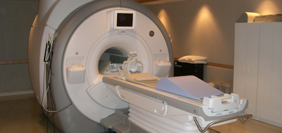
MRI scan uses a large magnet, radio waves and a computer to create a detailed image of person’s internal organs and structures. This is different from a CT scan where x-rays (radiation) is used to produce an image. MRI scan typically takes 45 minutes of actual scanning time to carry out MpMRI scan of prostate compared to few seconds to scan the whole body in most modern CT scanners.
MRI scans are considered harmless and do not have side effects. Most people tolerate MRI dye (gadolinium) very well and allergic reaction are relatively rare.
Some common problems to carry out MRI scan are:
- Claustrophobia – Some people find lying inside closed, confined space of an MRI scanner very daunting and are not able to complete the scan. This can sometimes be overcome by giving small dosage of sedation.
- Movement – During the scanning process, it is very important to lie very still for the entire during actual scanning. Though most people are able to cooperate reasonably well with this process, certain patients are unable to do so due to various reasons
- Hip Replacements – Though MRI scan be carried out in individuals with hip replacements, some of the sequences are very badly affected due to the presence of metal in the imaging field.
- Heart related implants – Patients with pacemakers and other cardiac implants and valves are generally not considered unless these objects are specifically MRI safe.
- Brain aneurysm clips and ear implants – These are other things to take into account and are not usually considered safe unless specified.
What is an MpMRI(Multiparametric MRI) scan?
Multiparametric scan is primarily MRI scan with additional functional sequences added on top of the standard sequences that are regularly used. We follow the guidance and protocol as specified by the European society of Uro-genital Radiologists (ESUR)
The two basic properties of cancer areas that are assessed here are
- tightly packed cells (Diffusion weighted imaging – DWI)
- increased blood supply (Dynamic contrast imaging – DCE)
Information from these sequences are combined with the information from all other sequences to grade the degree of suspicion in each area of prostate.
Both these sequences are severely affected in patients with artificial hips. However, sometime enough helpful information is obtained to aid the diagnosis.
Not all scanners are good enough to carry out these scans. The quality of scans will depend on the strength of the magnetic field, make of the scanners, age of the scanners and in general experience of the professionals carrying out these scans.
Can the scan be wrongly positive or negative?
Yes, the scans can be both wrongly positive or negative. Though majority of the times the scans correctly identifies the suspicious areas, it can be falsely positive or falsely negative.
According to PROMIS trial, it correctly identifies 93 out of 100 clinically significant cancers. This is much better than the conventional TRUS biopsy which identifies 48 out of 100 such cancers. However, when it identifies tumour it is correct only 41% of patients.
In our opinion, if the scan report is analysed in close conjunction with all the other information, accuracy of the scan is better. Moreover, the number of false positives decreases with experience of the professionals acquiring the images and the radiology doctors (radiologists) reporting the scan.
What happens after the scan?
The scan images are reported by skilled radiologist and the report is provided in text as well as a map to be seen by the referrer, usually a urologist. General practitioners with experience of the process may be able to make a judgement and refer to urologist as required.
Based on the report, your specialist will discuss with you, whether you need a biopsy and how this will be carried out.
What does the report usually say?
The report is provided in a standard format called PIRAD System as specified by ESUR. PIRADS stands for Prostate Imaging Reporting and Data System. Depending on the appearance of a specific area, a PIRADS score is provided which ranges between 1 and 5, 1 being probably benign and 5 being highly suspicious for malignancy.
PIRADS 1 – very low (clinically significant cancer is highly unlikely to be present)
PIRADS 2 – low (Clinically significant cancer is unlikely to be present)
PIRADS 3 – Intermediate (the presence of clinically significant cancer is equivocal)
PIRADS 4 – High (Clinically significant cancer is likely to be present)
PIRADS 5 –Very High (Clinically significant cancer is highly likely to be present)
If there are multiple areas that appear suspicious, each of these areas are scored and overall score is also provided.
As mentioned above, a negative scan does not completely exclude the tumour and positive scan does not always mean that it is cancer.
Final diagnosis has to be based on the biopsy and subsequent histology.